English
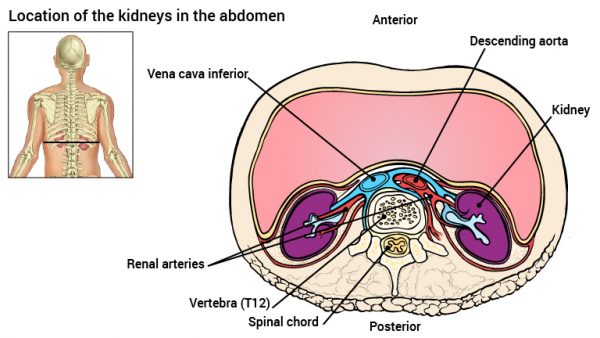
The body is equipped with two kidneys located in the posterior of the abdominal cavity at the level of the vertebrae T12 to L3 and are somewhat protected by the lower rib. The kidneys receive their blood supply from the aorta via the renal artery.
The kidneys are covered by three layers of supporting tissue. Outermost is the connective tissue that anchors the kidney in its location in the abdomen. The next layer is fatty tissue that protects the kidneys against physical injury and beneath this is a capsule composed again connective tissue that contains the main structures of the kidney.
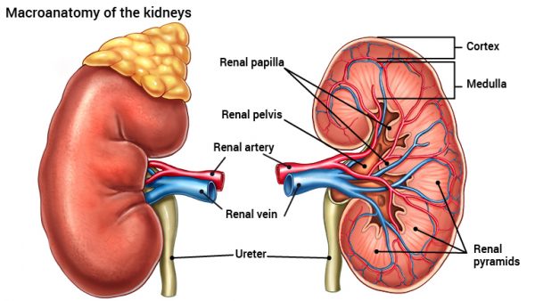
Directly beneath this connective tissue capsule, is the renal cortex and the inner layer of the kidney is the renal medulla. The gross structure of the medulla is comprised of eight pyramids. Each pyramid, contains thousands of the smallest structural and functional unit of the kidney, the nephron. Right in the centre of the kidneys are the papillae of each pyramid. These papillae act as a funnel and lead the filtered fluid to be excreted into the kidney pelvis. The renal pelvis is itself a ‘funnel and pipe’ that finally forms the ureter. The ureter channels the urine into the bladder where it is stored prior to excretion.
The kidneys receive under normal conditions about 25% of the cardiac output, i.e. on average a total of about 1.2 litres of blood per minute.
The autonomic nervous system is involved in the regulation of renal urine production as it is the sympathetic neurons that regulate renal blood supply.